Positron Emission Tomography (PET)
Positron emission tomography (PET) is a test that uses a special type of camera and a tracer (radioactive substance) to look at organs in the body. The tracer usually is a special form of a substance (such as glucose). It collects in cells that use a lot of energy, such as cancer cells.
During the test, the tracer liquid is typically put into a vein (usually in the arm), but sometimes it may be inhaled. The tracer moves through your body, where much of it collects in the specific organ or tissue. The tracer gives off tiny positively charged particles (positrons). The camera records the positrons and turns the recording into pictures on a computer.
PET scan pictures do not show as much detail as computed tomography (CT) scans or magnetic resonance imaging (MRI). This is because the pictures show only the location of the tracer. The PET picture may be matched with those from a CT scan to show more detailed information about where the tracer is located.
A PET scan is often used to evaluate cancer, check blood flow, or see how organs are working.
Why It Is Done
A PET scan is done to:
- Study the brain's blood flow and metabolic activity. A PET scan can help a doctor find nervous system problems, such as Parkinson's disease, multiple sclerosis, transient ischemic attack (TIA), amyotrophic lateral sclerosis (ALS), Huntington's disease, stroke, and schizophrenia.
- Find changes in the brain that may cause epilepsy.
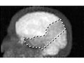
- Evaluate the extent of some cancers, especially lymphoma or cancers of the head and neck, brain, lung, colon, or prostate. In its early stages, cancer may show up more clearly on a PET scan than on a CT scan or an MRI.
- Determine whether a growth in an organ or in tissue is likely to be cancer, such as a growth in lung tissue.
- See how advanced a cancer is and whether it has spread to another area of the body (metastasized). Both CT and PET scans are often needed to evaluate cancer.
- Help a doctor choose the best treatment for cancer or to see how well treatment is working. PET scans may also be done to see whether surgery can be done to remove a tumor.
- Help diagnose Alzheimer's disease when the symptoms are not clear or when a person has dementia symptoms at a young age (usually younger than 65). This is called amyloid imaging.
- Find poor blood flow to the heart, which may mean coronary artery disease.
- Find damaged heart tissue, especially after a heart attack.
- Help choose the best treatment, such as coronary artery bypass graft surgery, for a person with heart disease.
How To Prepare
- Tell your doctor ALL the medicines, vitamins, supplements, and herbal remedies you take. Some may increase the risk of problems during your test. Your doctor will tell you if you should stop taking any of them before the test and how soon to do it.
- Tell your doctor if you have diabetes. If you take medicine to control diabetes, you may need to take less than your normal dose. Talk with your doctor about how much medicine you should take.
- Tell your doctor if you are or might be pregnant.
- If you are breastfeeding, you may want to pump enough breast milk before the test to get through 1 to 2 days of feeding. The radioactive tracer used in this test can get into your breast milk and is not good for the baby.
- You may not be able to eat or drink for at least 4 hours before some PET scans. Ask your doctor when or if you need to fast before the test.
- Your doctor will let you know if you should avoid smoking or avoid drinking caffeine or alcohol for 24 hours before this test.
- Your doctor will also let you know if you should avoid exercise or other strenuous activity for any period of time before this test.
- Tell your doctor if you get nervous in tight spaces. You may get a medicine to help you relax. If you think you'll get this medicine, be sure you have someone to take you home.
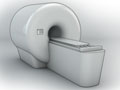
How It Is Done
A PET scan is done in a hospital nuclear medicine department or at a special PET center. You will lie on a table that is hooked to a large scanner, camera, and computer.
During the test
The radioactive tracer is usually given in a vein (I.V.). You may need to wait 30 to 60 minutes for the tracer to move through your body. During this time, you may need to avoid moving and talking.
The PET scanner, which is shaped like a doughnut, moves around you. The scanned pictures are sent to a computer screen so your doctor can see them. Many scans are done to make a series of pictures. It is very important to lie still while each scan is being done. At some medical centers, a CT scan will be done at the same time.
For a PET scan of the brain, you will lie on a bed. You may be asked to read, name letters, or tell a story, depending on whether speech, reasoning, or memory is being tested. During the scan, you may be given earplugs and a blindfold (if you do not need to read during the test) to wear for your comfort.
If you are having a PET scan of your heart, electrodes for an electrocardiogram (EKG, ECG) will be put on your body.
During the test, you may be alone in the scanner room. But the technologist will watch you through a window, and you'll be able to talk back and forth.
How long the test takes
The test will take 1 to 3 hours.
How It Feels
You will not feel pain during the test. The table you lie on may be hard and the room may be cool. It may be difficult to lie still during the test.
If you are getting the tracer through an I.V., you may feel a quick sting or pinch when the I.V. is put in your arm. The tracer is unlikely to cause any side effects. If you don't feel well during or after the test, tell the person who is doing the test.
You may feel nervous inside the PET scanner.
Risks
Allergic reactions to the tracer are very rare.
In rare cases, some soreness or swelling may develop at the I.V. site where the radioactive tracer was put in. Apply a moist, warm compress to your arm.
Anytime you're exposed to radiation, there's a small chance of damage to cells or tissue. That's the case even with the low-level radioactive tracer used for this test. But the chance of damage is very low compared with the benefits of the test.
Results
The radiologist may discuss preliminary results of the PET scan with you right after the test. Complete results are usually available in 1 to 2 days.
Normal
- Blood flow is normal and organs are working well. The flow and pattern of the tracer shows normal distribution in the body.
Abnormal
- Heart:
- Decreased blood flow and increased glucose metabolism may show that the blood vessels are narrowed or blocked. This may mean coronary artery disease (CAD) is present.
- Decreased blood flow and glucose metabolism may mean that heart tissue is scarred and damaged. This may be from a heart attack.
- Brain:
- Areas of increased glucose metabolism or lower oxygen use and blood flow may mean you have epilepsy.
- Decreased oxygen use and blood flow may mean a stroke has occurred.
- Decreased glucose metabolism may mean a form of dementia. Dementia may be caused by Parkinson's disease or Huntington disease. Or it may be from mental illness, such as schizophrenia.
- Patterns of blood flow and oxygen use that are not normal may mean a brain tumor is present.
- A special test (called amyloid imaging) may show signs of Alzheimer's disease.
- Tumor detection:
- Areas of increased glucose metabolism may mean a tumor is present.
Credits
Current as of: March 26, 2025
Author: Ignite Healthwise, LLC Staff
Clinical Review Board
All Ignite Healthwise, LLC education is reviewed by a team that includes physicians, nurses, advanced practitioners, registered dieticians, and other healthcare professionals.
Current as of: March 26, 2025
Author: Ignite Healthwise, LLC Staff
Clinical Review Board
All Ignite Healthwise, LLC education is reviewed by a team that includes physicians, nurses, advanced practitioners, registered dieticians, and other healthcare professionals.